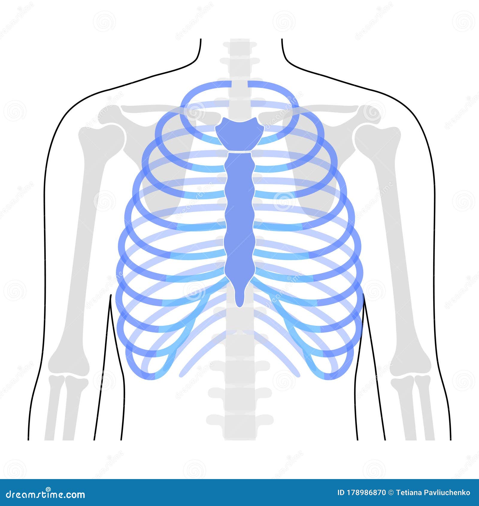
If you go and search for a picture of human rib cage right now, you're probably going to see the same sterile, perfectly symmetrical white bones against a blue or black background. It looks neat. It looks tidy. Honestly, it's also kinda misleading. Real human anatomy isn't a plastic model from a high school biology classroom, and the way our ribs actually sit in our bodies is way more dynamic—and crowded—than most stock photos suggest.
You’ve got 24 ribs. Most people do, anyway.
But here’s the thing: about one in 200 to 500 people is born with an extra one called a cervical rib. It sits right above the first rib. If you’re looking at a standard medical diagram, you’d never know that exists, yet for the person holding that X-ray, it’s a massive deal because it can cause thoracic outlet syndrome. This is why understanding what you’re looking at in an anatomical image matters more than just "bone counting."
Why Your Perspective Matters When Viewing a Picture of Human Rib Cage
The angle of the image changes everything. When you look at a frontal view (anterior), the rib cage looks like a flat shield. It isn’t. If you saw a "bird's eye" view looking down through the neck, you'd see the ribs actually form a heart-shaped or kidney-shaped opening.
Most people think the ribs are just there to protect the heart and lungs. That’s the "cage" part of the name. But they’re also a massive mechanical engine. Every time you inhale, those ribs move like bucket handles. They swing up and out. If they were as rigid as they look in a static picture of human rib cage, you wouldn't be able to take a single breath.
Dr. Alice Roberts, a renowned anatomist, often points out how the evolution of our rib cage reflects our transition to walking upright. Our thorax is actually quite flat from front to back compared to other primates. This shifts our center of gravity, making it easier for us to balance on two legs. When you look at a lateral (side) photo, you can see that subtle "S" curve of the spine interacting with the ribs. It’s a masterpiece of engineering, really.
The Mystery of the Floating Ribs
Look at the very bottom of any decent anatomical illustration. You’ll see two pairs of ribs that don’t wrap all the way around to the front. They just... hang there. These are the 11th and 12th ribs, better known as the floating ribs.
Why are they there?
They provide a bit of protection for the kidneys, but their main "job" is to stay out of the way. If those ribs attached to the sternum like the others, you wouldn’t be able to twist your torso or bend over nearly as well. They allow for the flexibility of the lower back. In some historical medical texts—and honestly, in some weird modern myths—people claim you can have these removed for a smaller waist. While "rib removal" surgery does exist, it’s incredibly rare and carries huge risks, including potential organ prolapse.
The Cartilage Gap: What Photos Usually Miss
One of the biggest issues with a standard picture of human rib cage is that it often ignores the costal cartilage. In a real body, the bony part of the rib doesn't touch the breastbone (sternum). Instead, there’s a thick, rubbery bridge of hyaline cartilage.
- This cartilage is what allows the chest to expand.
- It’s also why CPR works. If the chest were solid bone, a paramedic would just snap your ribs immediately (though, to be fair, ribs often do crack during high-quality CPR anyway).
- In older adults, this cartilage can actually start to calcify, turning into bone. This is why breathing often feels "stiffer" as people age.
If you’re looking at an image for artistic reference, pay attention to the "subcostal angle." That’s the upside-down 'V' shape where the ribs meet at the bottom of the sternum. In some people, it’s narrow; in others, it’s wide. This variation is why some people look "barrel-chested" while others look lean, regardless of how much they work out.
Identifying Fractures and Anomalies in Medical Imagery
If you’re looking at a picture of human rib cage because you’re worried about a personal injury, keep in mind that rib fractures are notoriously hard to spot on standard X-rays. Radiologists often look for the "fatty tissue sign" or a disruption in the smooth white line of the bone’s cortex.
A "simple" break might look like a tiny hair-thin crack. But a "flail chest"—which is as scary as it sounds—occurs when multiple ribs break in two places. This creates a free-floating segment of the chest wall. In a clinical photo or CT scan of this, you’d see the chest wall moving inward when the patient tries to breathe out. It’s a paradox that defies how we think the "cage" should work.
Real-World Variations You Should Know About
Not every rib cage is "normal." Pectus excavatum is a condition where the breastbone sinks into the chest. In a picture of human rib cage with this condition, it looks like there’s a bowl-shaped hollow in the center of the torso. On the flip side, Pectus carinatum causes the sternum to protrude like a bird’s breast.
These aren't just "cosmetic" differences. They can actually squeeze the heart or limit lung capacity. When medical illustrators create images, they usually aim for the "average," but the average person doesn't actually exist. Everyone has a slightly different curvature.
How to Read a Rib Cage Image Like a Pro
If you want to actually understand what you're looking at, follow the "rule of segments."
- The True Ribs (1-7): These attach directly to the sternum. They are the most stable.
- The False Ribs (8-10): These attach to the cartilage of the rib above them. They create that flared look at the bottom of the ribs.
- The Floating Ribs (11-12): No attachment to the front.
When you see a 3D render or a high-quality photograph, look at the spaces between the ribs. These are the intercostal spaces. They aren't empty. They are packed with three layers of muscles, plus an artery, a vein, and a nerve. This "neurovascular bundle" sits right in a groove on the underside of each rib. This is why doctors always insert chest tubes above a rib, never below it—you don't want to hit that nerve or bleed out from the artery.
Using Images for Study or Art
For artists, the rib cage is the "chassis" of the figure. If you get the tilt of the rib cage wrong in relation to the pelvis, the whole drawing falls apart. Professional artists often use a "simplified" picture of human rib cage that looks more like an egg or a bell. This helps in visualizing the volume of the chest before adding the pectoral muscles or the latissimus dorsi.
For students, don't just look at one image. Look at a "coronal" slice (from the front), a "sagittal" slice (from the side), and a "transverse" slice (from the top). This 3D understanding is the only way to truly grasp how the heart sits nestled behind the sternum, slightly to the left, protected by this bony armor.
Actionable Next Steps for Further Research
If you are trying to find the most accurate picture of human rib cage for medical or educational purposes, skip the basic Google Image search and go straight to these high-fidelity sources:
- The Visible Human Project: This provides cross-sectional images of a real human body. It’s the "gold standard" for seeing how the ribs interact with organs.
- Kenhub or TeachMeAnatomy: These sites offer peer-reviewed illustrations that clearly distinguish between bone and costal cartilage.
- Radiopaedia: If you want to see what ribs look like in real-world scenarios (fractures, pathologies, and variations), this is a database of actual clinical X-rays and CT scans used by radiologists.
- BioDigital Human: A free 3D platform that lets you rotate the rib cage and strip away layers of muscle to see the bone structure in a 360-degree space.
Don't settle for the first stylized drawing you see. Real anatomy is messy, asymmetrical, and incredibly functional. Whether you’re studying for a kinesiology exam or just trying to figure out why your side hurts after a cough, understanding the nuances of the thoracic cage is the first step toward body literacy.
Focus on the connection points—the sternum in the front and the thoracic vertebrae in the back. That's where the real "action" happens. Once you see the rib cage as a moving, living part of your respiratory system rather than just a static cage, those diagrams start to make a lot more sense.