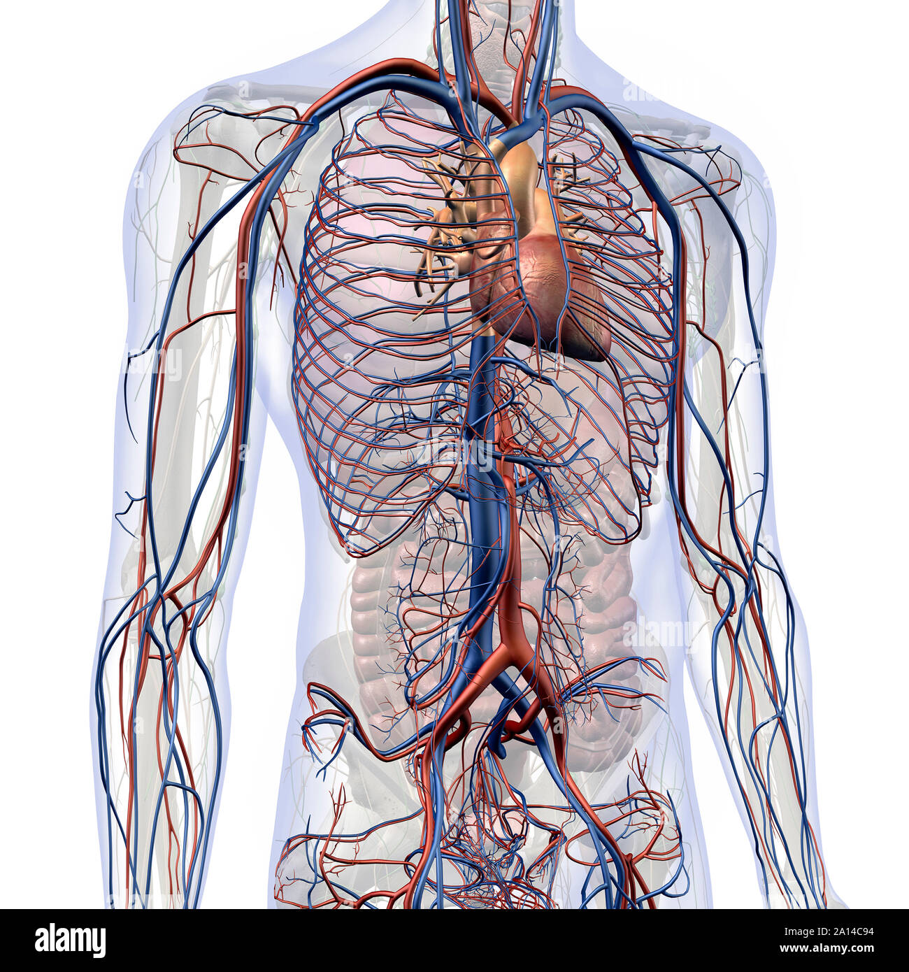
Honestly, it’s kind of wild how much we simplify the human body when we’re in school. Most people think "chest" and immediately jump to lungs or maybe the heart, but when we look at female chest anatomy organs, we’re dealing with a layered, multi-system setup that manages everything from your literal survival to the immune system and reproductive biology. It’s not just a cage for your breathing bits.
You’ve got the superficial stuff like the breasts, which are technically modified sweat glands (weird, right?), and then you dive deep into the thoracic cavity where the heavy hitters live. We are talking about a dense neighborhood of tissue, bone, and vascular highways.
Understanding this isn't just for medical students. It’s for anyone who wants to know why a certain pain feels the way it does or why the "chest" is actually the most complex real estate in the body.
The Breasts: More Than Just Soft Tissue
People usually call them breasts and leave it at that, but the internal mechanics are fascinatingly complex. The mammary glands are the stars here. These aren't just solid lumps; they are composed of 15 to 20 lobes of glandular tissue, arranged sort of like the petals of a daisy.
Each lobe contains smaller lobules where milk is actually produced. This whole system is drained by lactiferous ducts that lead to the nipple. But here is what most people miss: the amount of fat in the breast determines the size, but it has absolutely zero impact on how much milk those glandular organs can produce. A "small" breast can have just as much functional glandular tissue as a "large" one.
Between these lobes are Cooper’s ligaments. Think of them as the internal suspension cables. They’re these tough, fibrous bands of connective tissue that hook onto the skin and the underlying fascia of the chest muscles. Over time, gravity and the breakdown of collagen mean these cables stretch. It’s just physics.
The Thoracic Cage: The Body’s Armor
Underneath the soft tissue lies the rib cage, or the thoracic cage. It’s a flexible vault. You’ve got 12 pairs of ribs, and while there’s an old myth that women have more ribs than men, that is total nonsense. Everyone (mostly) has 24.
The first seven pairs are "true ribs" because they connect directly to the sternum via costal cartilage. Then you’ve got the "false ribs" and those two "floating ribs" at the bottom that just hang out without attaching to the front at all. This cage does more than protect; it’s a pump. Without the intercostal muscles between those ribs pulling the cage up and out, you wouldn't be able to create the pressure change needed to draw air into your lungs.
The Lungs and the Pleural Space
Inside that cage, the lungs take up the most volume. They aren't equal, though. The right lung has three lobes, but the left lung only has two. Why? Because the heart needs a place to sit. The "cardiac notch" is a little indent in the left lung specifically designed to make room for the heart’s apex.
What’s really cool is the pleura. This is a thin, double-layered membrane. One layer sticks to the lung, the other sticks to the chest wall. In between is a tiny bit of fluid. This setup creates surface tension—basically like two wet pieces of glass stuck together. This tension is the only reason your lungs don't just collapse into a pile of tissue every time you exhale.
The Heart: The Central Engine
Nestled in the mediastinum—the central compartment of the thoracic cavity—is the heart. In the context of female chest anatomy organs, the heart is often smaller than a male's, and it beats slightly faster to compensate for its size.
It sits slightly to the left of the midline. It’s wrapped in the pericardium, a tough sac that keeps it in place and prevents it from over-expanding if your blood pressure spikes. Doctors like Dr. Nanette Wenger, a pioneer in women's cardiology, have spent decades pointing out that because of this anatomical positioning and hormonal differences, women’s "chest pain" often feels different than men’s. It’s not always a "crushing weight"; sometimes it’s just a dull ache or pressure because of how the nerves in the thoracic cavity are bundled.
The Thymus: The Forgotten Immune Organ
Here is one nobody talks about. Directly behind your sternum and in front of your heart sits the thymus. In kids, it’s huge. In adults, it starts to shrink and turn into fat—a process called involution.
But don't ignore it. The thymus is the "schoolhouse" for T-cells. It’s where your immune system learns to distinguish between "you" and "not you." Even as an adult, having a healthy thoracic environment means protecting the remnants of this gland. It’s basically the headquarters for your body’s internal security force during your formative years.
The Esophagus and the Great Vessels
Running right down the middle, behind the trachea, is the esophagus. It’s a muscular tube that stays collapsed until you swallow something. It shares space with the aorta—the massive "garden hose" artery that carries oxygenated blood out of the heart.
The proximity here is why "heartburn" feels like it's coming from your heart. The esophagus is literally inches away from the cardiac tissue. When stomach acid irritates the esophageal lining, the nerves in your chest can't always tell exactly where the fire is coming from.
Why This Anatomy Matters for Health
Knowing the layout changes how you handle your health. For example, the axillary lymph nodes—located in the upper outer quadrant of the breast and up into the armpit—are the primary drainage system for the chest. This is why when doctors do a breast exam, they always feel up into the underarm. They’re checking the "filters" of the chest.
Practical Steps for Chest Health
- Posture and Expansion: Because the ribs and intercostals are mechanical, "hunching" over a phone actually restricts the volume your lungs can intake. Practice "diaphragmatic breathing" to keep the costal cartilage flexible.
- Self-Exams: Don't just look for "lumps" in the breast tissue. Look for changes in the skin texture or nipple discharge. Remember the "tail of Spence"—the part of the breast tissue that extends toward the armpit.
- Cardiovascular Awareness: Since the female heart is anatomically different in its vessel structure (often having smaller coronary arteries), regular aerobic exercise is non-negotiable for maintaining the elasticity of the thoracic aorta.
- Check the Sternum: If you feel pain right in the middle of your chest that hurts when you press on it, it might not be your heart or lungs at all. It could be costochondritis—inflammation of the cartilage connecting the ribs to the sternum.
Understanding your chest anatomy is basically like having a map of your own engine room. When you know where the parts are, you stop panicking at every "thump" and start knowing when to actually call a mechanic (or a doctor). Pay attention to the symmetry. Most of these organs are paired or centered. If something feels "off" on just one side, that’s usually your body’s way of flagging a specific system for review.
Stay aware of the lymph nodes under the collarbone as well. These supraclavicular nodes are often the first to swell if there's an infection or systemic issue within the chest cavity. Keeping an eye on these anatomical landmarks is your first line of defense in proactive wellness.
Actionable Insight: Set a recurring monthly reminder on your phone for a "Chest Health Check." Spend three minutes checking for lymph node tenderness under the arms and along the collarbone, and practice five deep, rib-expanding breaths to maintain thoracic mobility. This simple habit connects you to your internal anatomy and helps you catch subtle changes before they become major issues.